Skip to presentation

ACCSM Annual Scientific Meeting · 13 August 2026

Repositioning of the Breast Footprint in Revision Augmentation Using Total Implant Encapsulation with Synthetic Mesh.

Revision augmentation · total implant encapsulation · synthetic mesh

Dr Kishen Nara

MBBS (Monash) · FACCSM(Surg)

revaesthetic.com.au

White sand and turquoise water on the Tasmanian coast
Greetings from Tasmania

Plate II · Declaration

Disclosures

Declaration

No relevant financial relationships to disclose.

02

Plate III · The brief

Case at a glance

Patient age 56 years
Weight
66 kg
Height
178 cm
BMI
20.8
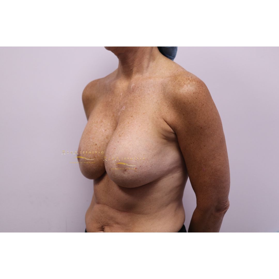
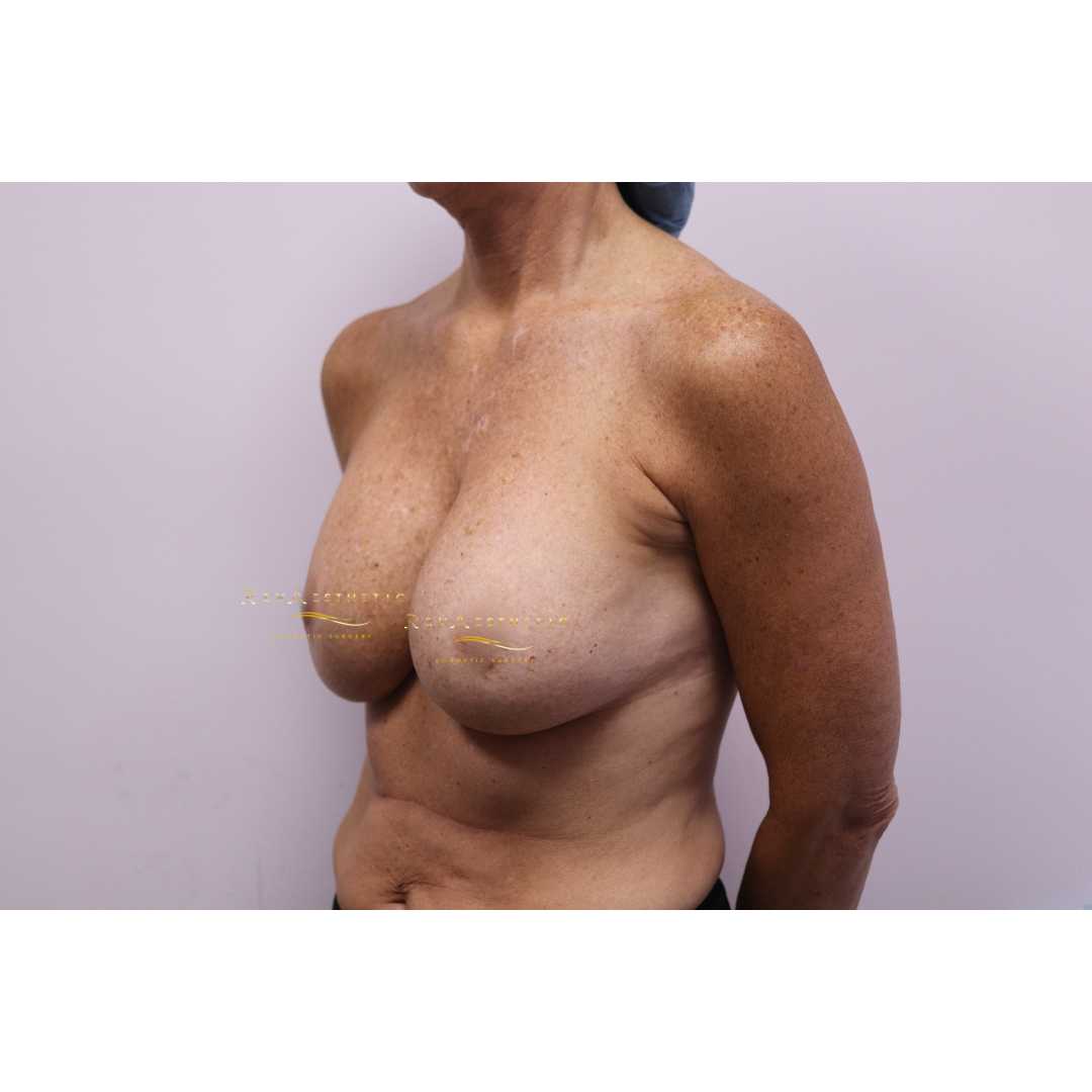
De-identified pre-operative left oblique clinical photograph
Left oblique
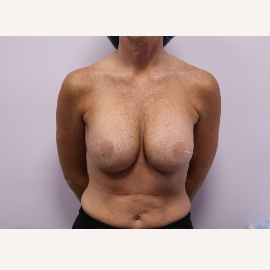
De-identified pre-operative AP clinical photograph
AP
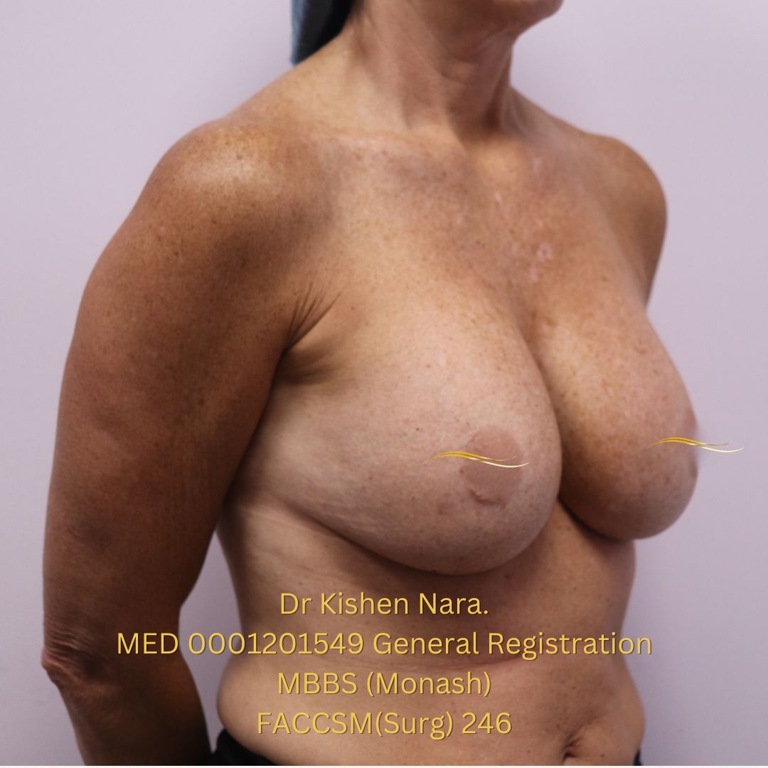
De-identified pre-operative right oblique clinical photograph
Right oblique

03

Plate IV · Desired direction

Patient goals

Up to675 ccdiscussed preoperatively

  • Larger, round, high-profile form
  • Greater upper-pole fullness
  • A higher breast position
  • No mastopexy · no new scars

04

Plate V · Starting position

Prior augmentation

2005

Primary procedure

Thailand · IMF incision · submuscular pocket

Unknown pre-operatively

Existing implant

Device details were unknown before surgery; estimated volume 350–380 cc.

Presenting concerns

Too small and less firm

Lower position with a deflated appearance.

05

Plate VI · Context

Relevant history

Obstetric
G3P3
Breast
Pre-gravid 10C · increased to DD while breastfeeding
General health
Fit, healthy, stable weight
Haematology
Iron-deficiency anaemia resolved after iron infusions
Smoking
Ex-smoker · ceased 2008
Other
Gluten intolerance

06

De-identified pre-operative AP clinical photograph
Pre-operative AP
De-identified pre-operative left oblique clinical photograph
Pre-operative left oblique

Plate VII · Examination

Clinical examination

4–5 mmsoft-tissue thickness overall
22 / 22.75 cmSN–N · R / L
Grade IIIright capsular contracture
13.75 / 14 cmbreast width · R / L
10.5 / 10 cmN–IMF · R / L
81 / 89 cmunderbust / overbust
  • Bilateral pseudoptosis
  • Waterfall deformity · left

07

Plate VIII · Clinical considerations

Can a larger implant be placed in a thin envelope while repositioning the footprint?

01

Precision pocket design

Define the new footprint while preserving the thin soft-tissue envelope.

02

Placement & support

Control implant position and reinforce the inferior and lateral pocket.

03

Coverage

Anticipate rippling and palpability with 4–5 mm tissue thickness.

04

Longevity

Plan for future lower-pole elongation and recurrent ptosis.

08

Plate IX · Decision architecture

Options discussed

  1. 01

    No intervention

    Accept the current volume and position.

  2. 02

    Defer

    Reconsider if priorities or tissue conditions changed.

  3. 03

    Removal & replacement

    Inferior mesh support.

  4. 04

    Selected pathway

    Removal & replacement

    Total encapsulation of the implant with mesh.

  5. 05

    Mastopexy

    Discussed and declined.

09

Plate X · Due diligence

Further pre-op consultations

01

MRI

Not completed due to claustrophobia

02

Imaging

Mammogram and ultrasound

03

Haematology

Iron deficiency treated and optimisation confirmed

04

Team review

Multidisciplinary consultation and operative planning

10

Plate XI · Consent

Risks of the selected operation

≈8%complications quoted
≈15%revision quoted

Figures recorded in this individual consent discussion.

  • Capsular contracture
  • Rupture or leak
  • Rippling
  • Malposition
  • BIA-ALCL
  • Palpability
  • Future ptosis
  • Further revision

Access strategy

Re-use of the IMF scars was discussed with the FACCSM(Surg) and RACS team to protect the thin-skin blood supply. Temporary indentation was anticipated.

11

Plate XII · Biomaterial selection

Three mesh generations

TIGR® Matrix Surgical Mesh · manufacturer introduction · first 70 seconds

Comparison of permanent synthetic mesh, acellular dermal matrix and synthetic resorbable mesh
Synthetic permanentPermanent polymer meshDurable support; permanent foreign material.
ADMBiologic tissue matrixRemodels after incorporation; donor material and cost.
Synthetic resorbableTemporary polymer scaffoldResorbs over time; long-term breast evidence remains limited.

Selection depends on tissue quality, pocket control, contamination risk, availability and intended duration of support.

[1–4] For TIGR, meaningful slow-fibre support and complete material resorption are distinct endpoints.

12

Hand-drawn sagittal breast anatomy showing green TIGR mesh closely surrounding an implant and fixed to the third rib
Operative pocket design after superior and lateral capsulotomy, with the TIGR mesh closely surrounding the implant and fixed to the third rib.
Superior + lateral capsulotomy Total implant encapsulation in TIGR Third rib

Plate XIII · Operative design

Build the pocket before choosing the volume.

  1. 01

    Submuscular explant and revision

  2. 02

    Superior and lateral capsulotomy

  3. 03

    Partial capsulectomy

  4. 04

    TIGR internal pocket reinforcement

  5. 05

    610 cc Polytech Même POLYtxt XHP implant

  6. 06

    Drains

13

Plate XIV · Operative sequence

Animated operative sequence

14

Plate XV · In theatre

The pocket selected the implant.

Sizers trialled510 · 590 · 610 · 650 · 675 cc
Final implant610 cc

Polytech Même POLYtxt · Round · XHP · 65 mm projection

TIGR Matrix 20 × 30 cm each side · drains placed.

Explant: 15-year-old 375 cc smooth Mentor moderate-profile implant.

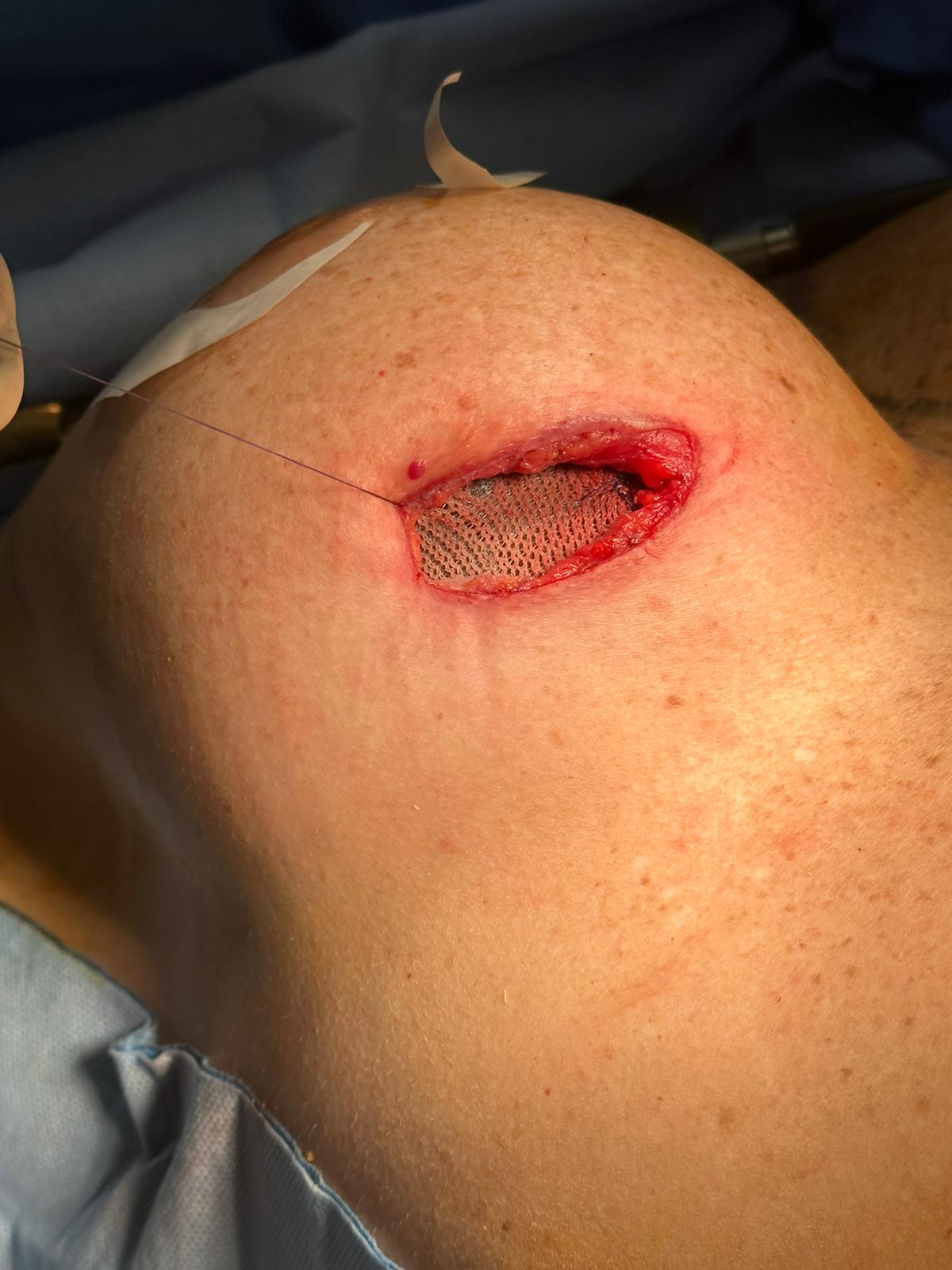
Intra-operative photograph showing TIGR mesh visible through the inframammary incision
Intra-operative · TIGR mesh in situ
Magnified intra-operative detail of TIGR mesh through the inframammary incision
Magnified detail · mesh at the IMF access point

15

Plate XVI · Follow-up

Post-operative course

Day 1

Happy

Some nausea

3 weeks

Firmer and symmetric

Wounds healed · shoulder movement good

11 months

Satisfied

Scars healed · tissue thickness slightly improved

≈3 years

Follow-up continued

Reviewed to approximately the 3-year mark

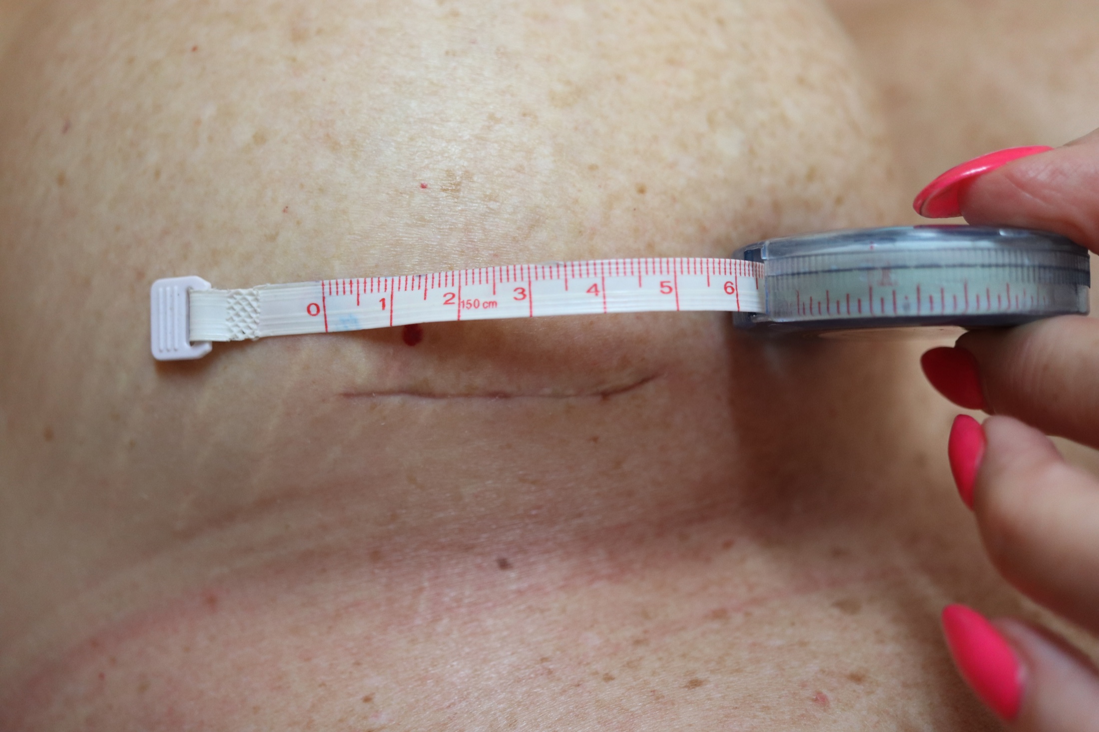
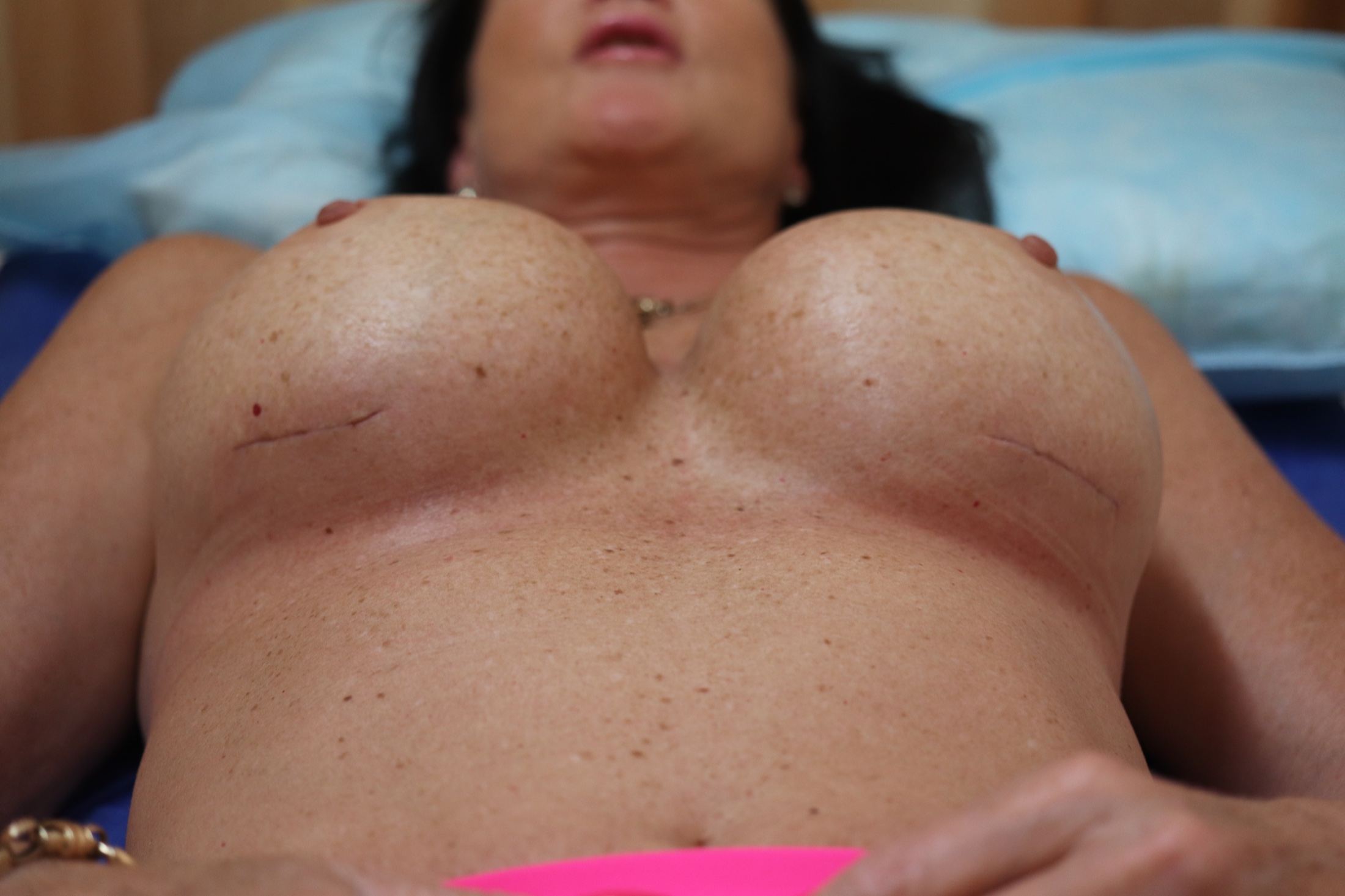
De-identified close-up photograph measuring an inframammary fold scar
IMF scar measurement · View 1
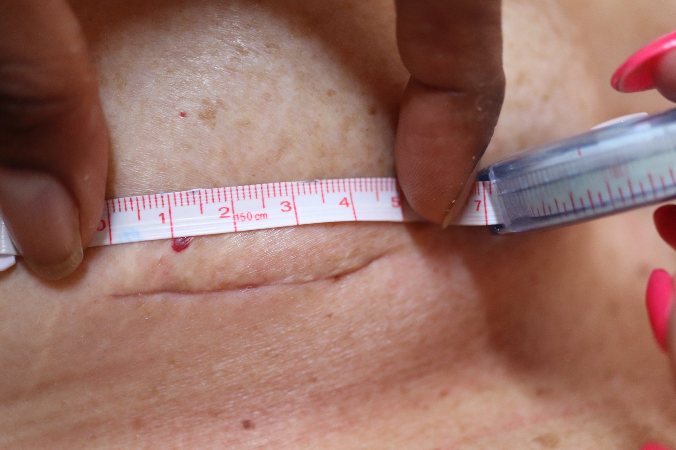
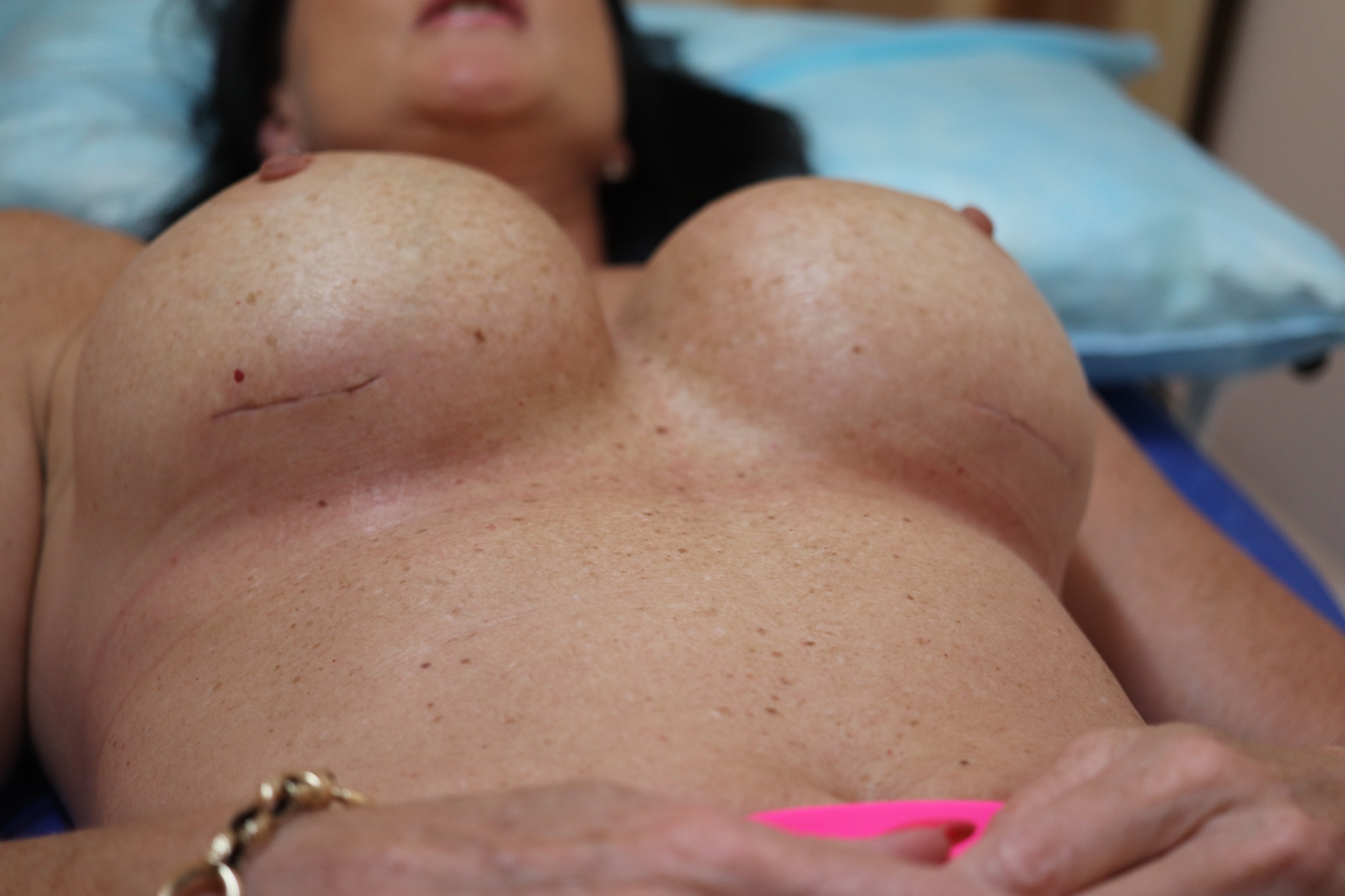
De-identified close-up photograph measuring an inframammary fold scar
IMF scar measurement · View 2
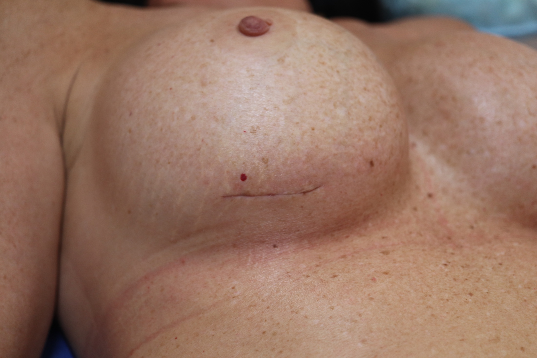
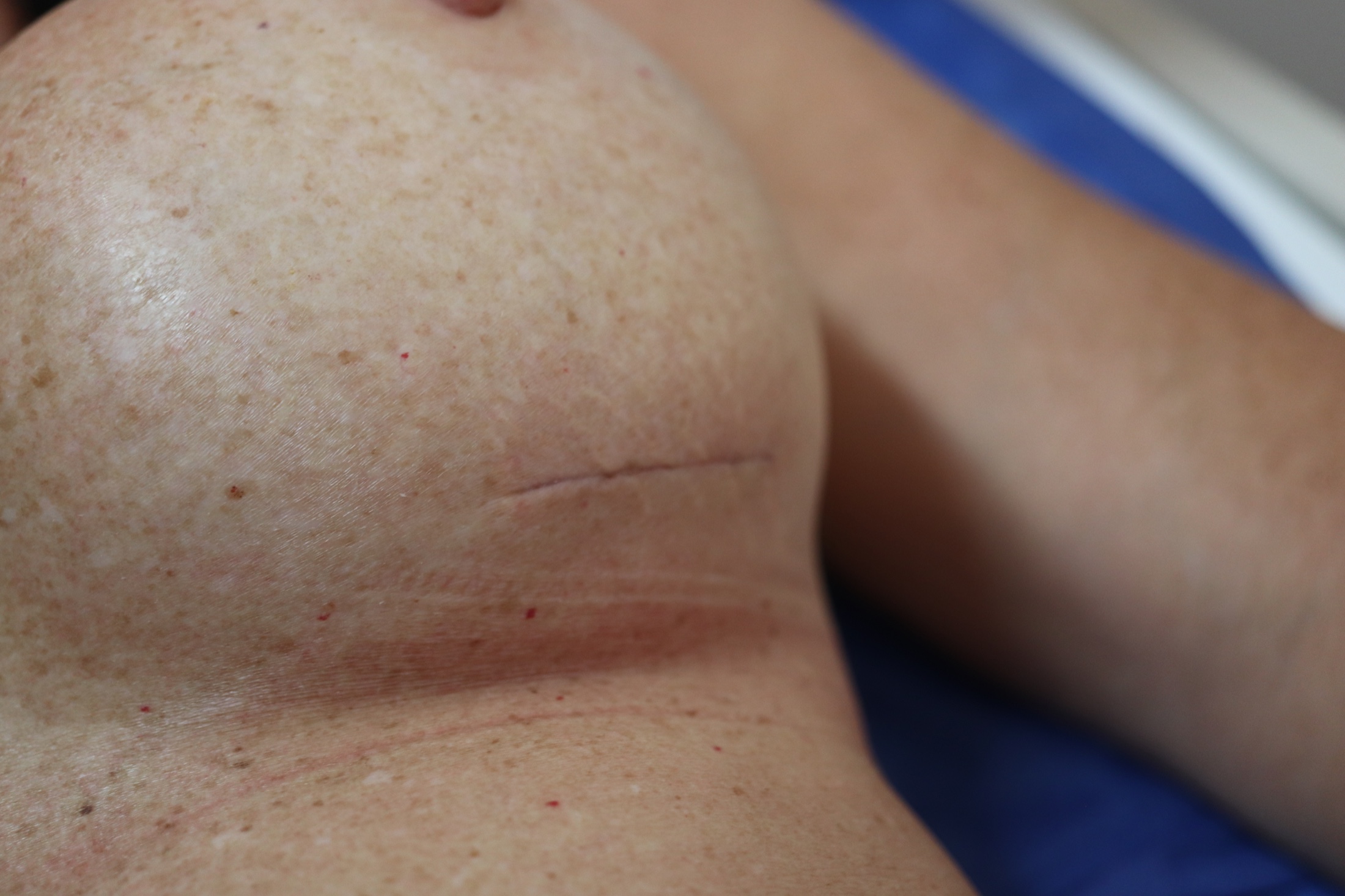
De-identified close-up photograph of a healed inframammary fold scar
Healed IMF scar · View 1
De-identified close-up photograph of a healed inframammary fold scar
Healed IMF scar · View 2

Single-case observation

16

Plate XVII · Clinical photography

Clinical follow-up series

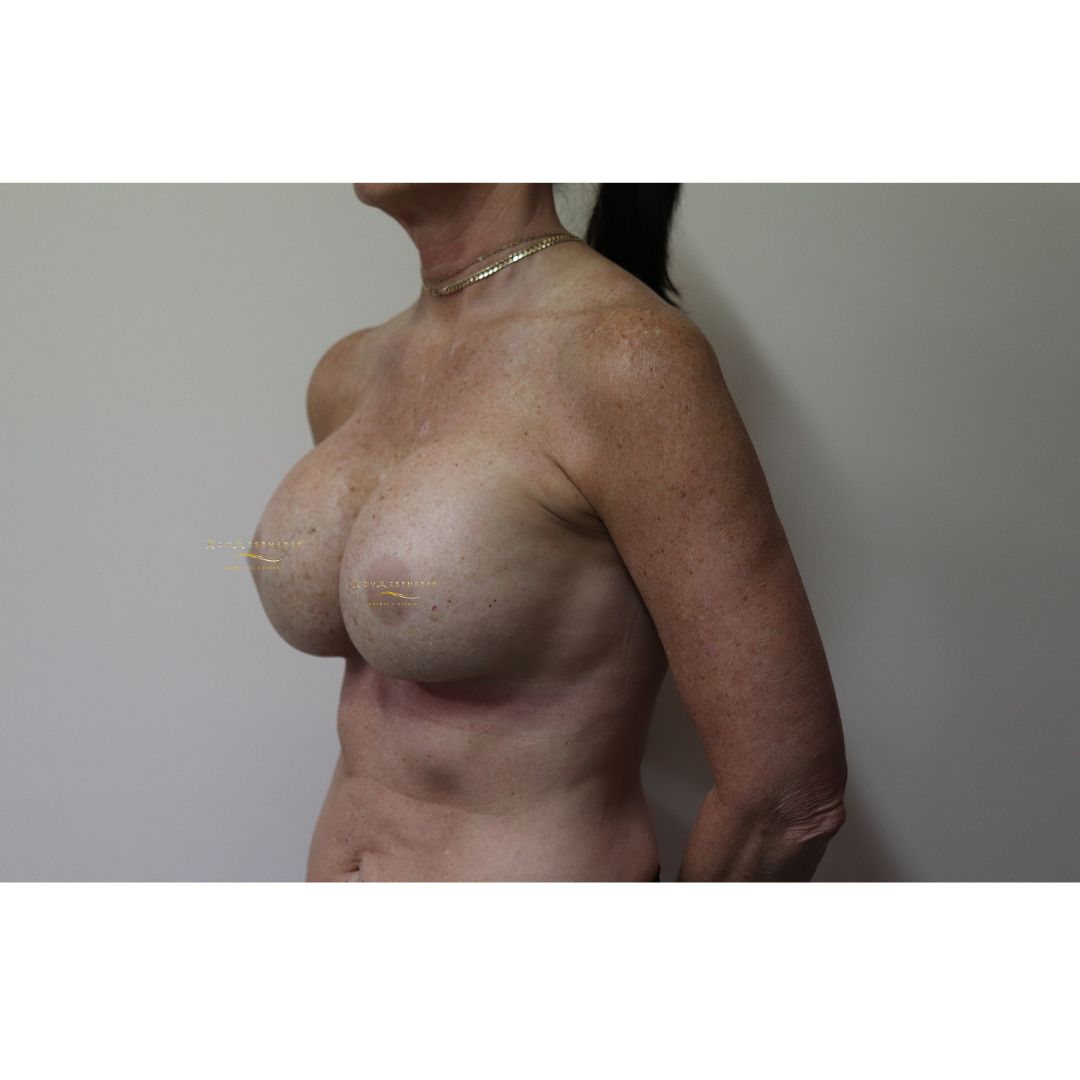
De-identified post-operative left oblique clinical photograph
Left oblique01
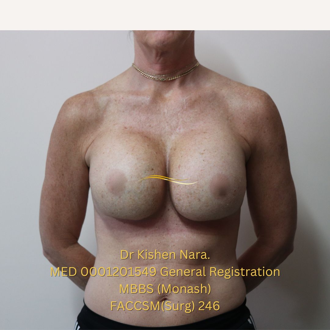
De-identified post-operative AP clinical photograph
AP02
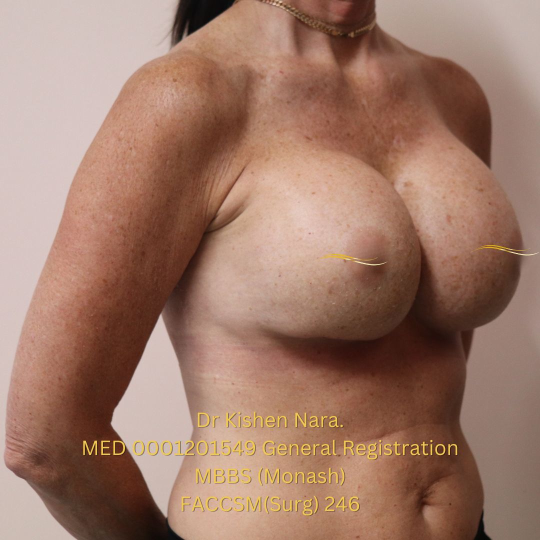
De-identified post-operative right oblique clinical photograph
Right oblique03
De-identified inferior bilateral follow-up view showing the inframammary fold scars
Supine frontal04
De-identified inferior bilateral follow-up view showing the breast footprint and inframammary fold scars
Inferior oblique05
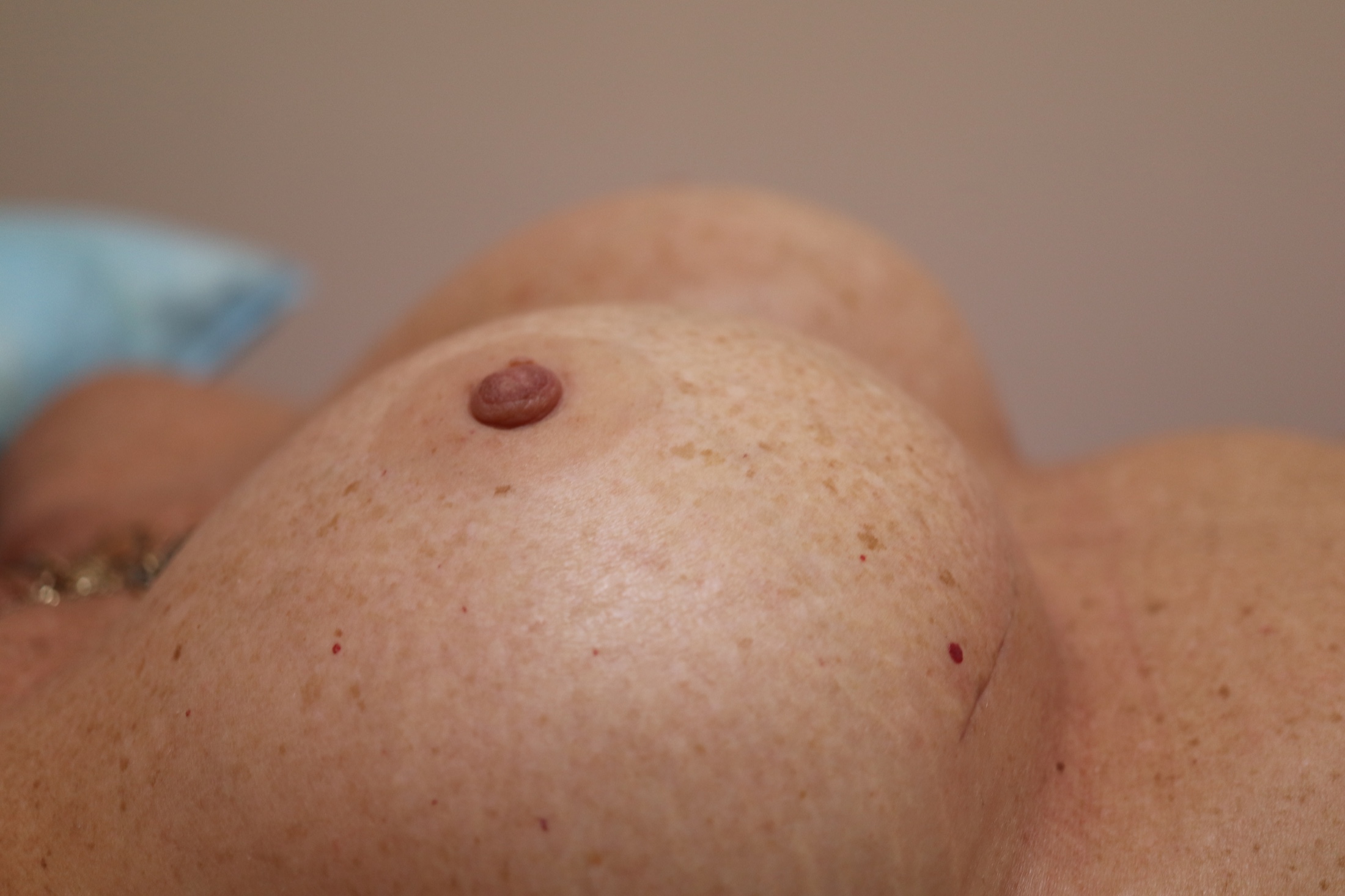
De-identified close-up follow-up view of breast contour and projection
Contour detail06
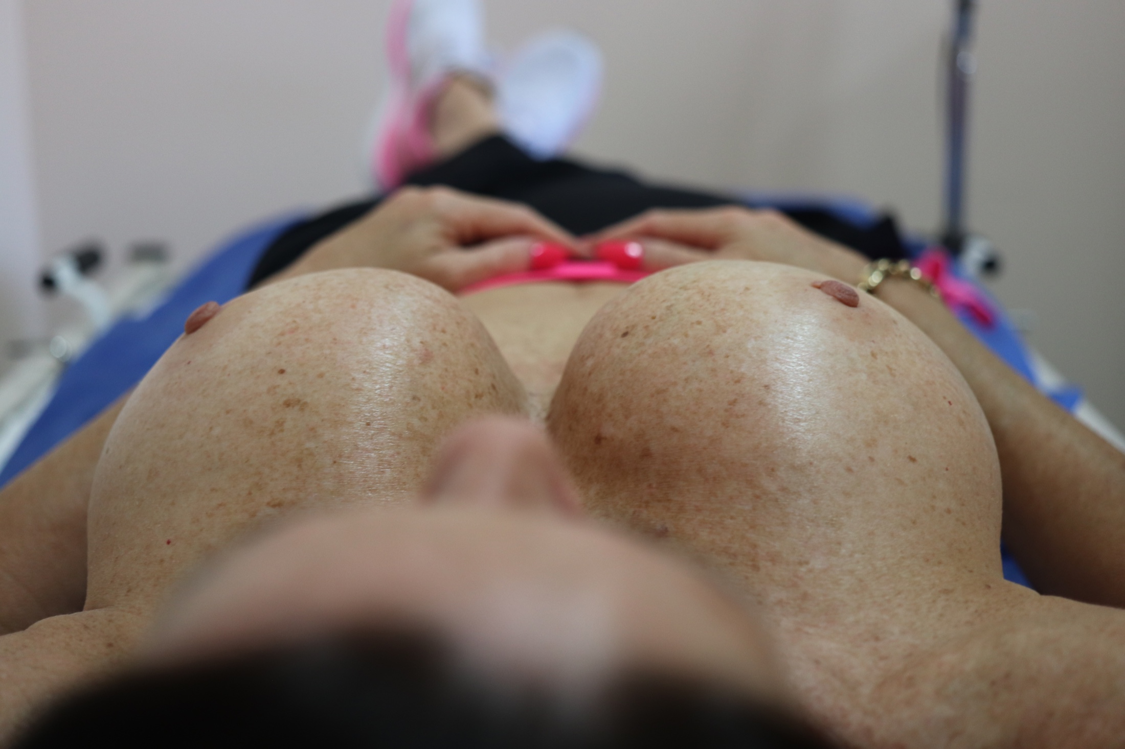
De-identified inferior bilateral follow-up view demonstrating breast footprint symmetry
Supine projection07
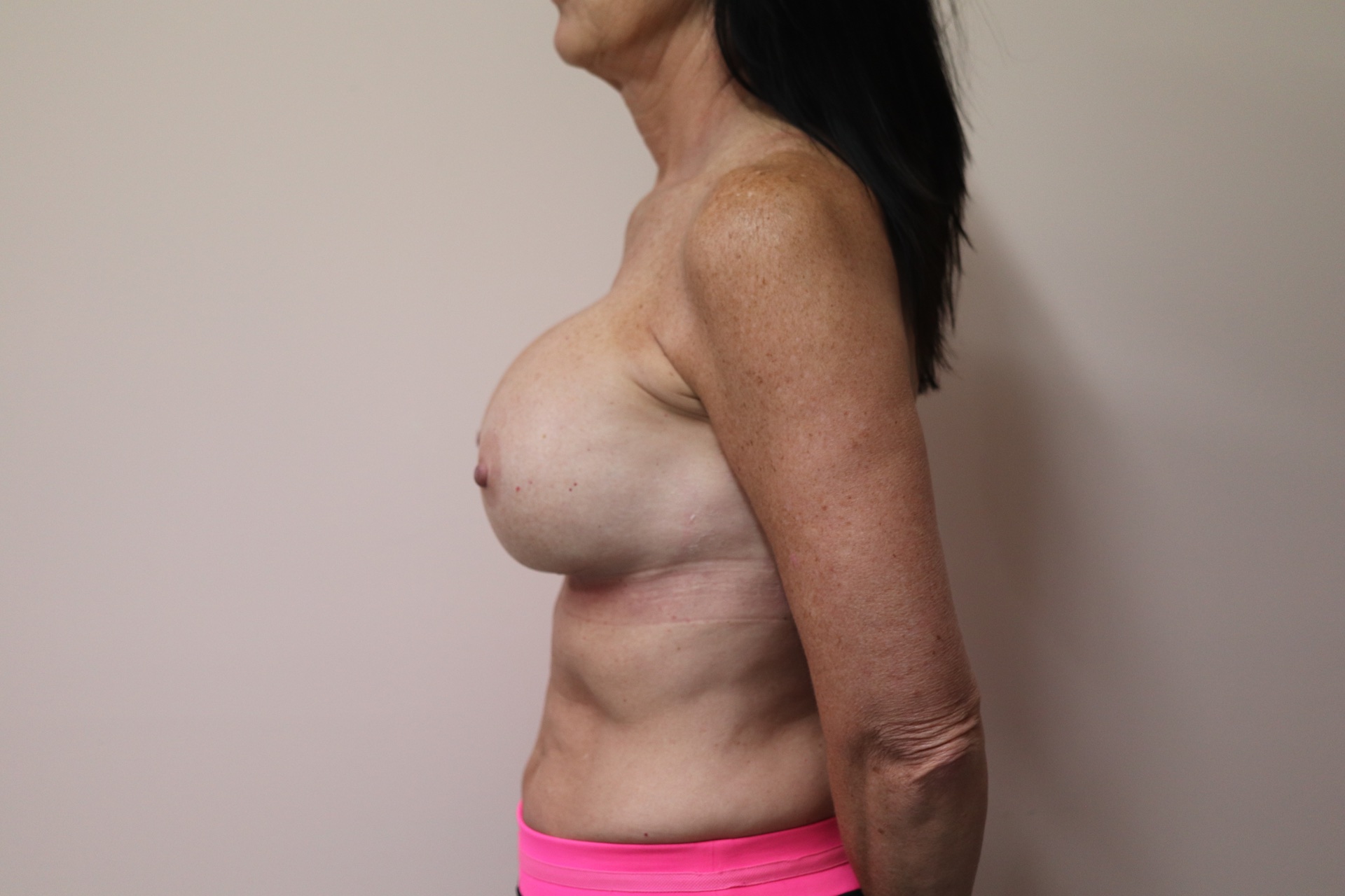
De-identified standing lateral post-operative clinical photograph
Standing lateral08

17

Plate XVIII · Before and after

Matched clinical views

Before first · same patient · individual results vary

Frontal AP

De-identified frontal pre-operative clinical photograph
Before01
De-identified frontal post-operative clinical photograph
After02

Left oblique Matched angle

De-identified left oblique pre-operative clinical photograph
Before03
De-identified left oblique post-operative clinical photograph
After04

Right oblique Matched angle

De-identified right oblique pre-operative clinical photograph
Before05
De-identified right oblique post-operative clinical photograph
After06

18

Plate XIX · Discussion

Four decisions carried the case

01

Reinforce selectively

Support the compromised lower pole and lateral pocket without leaving permanent polypropylene.

02

Control the pocket

Implant footprint, dissection and fixation remain primary; mesh is an adjunct.

03

Respect vascularity

Re-use existing scars deliberately when the envelope is thin and previously operated.

04

Align expectations

The no-lift constraint, future ptosis, palpability and revision risk must remain explicit.

Evidence context

Breast-specific evidence remains limited and largely observational; this case supports technical discussion, not a comparative effectiveness claim. [1–4]

19

Plate XX · Conclusion

What this case demonstrates

  1. 1

    Plan the footprint first. Implant volume, projection, pocket boundaries and tissue coverage must be planned as one problem.

  2. 2

    Mesh remains an adjunct. Total encapsulation supported pocket control; it did not replace precise dissection, fixation or implant selection.

  3. 3

    Interpret one case cautiously. At approximately 3 years, the repositioned footprint was maintained in this patient; broader conclusions require stronger evidence.

20

REVAESTHETIC

Discussion

Thank you.

Questions & discussion

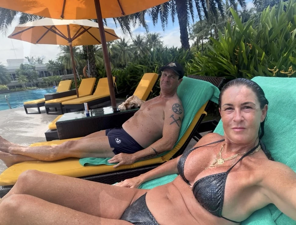
Patient enjoying an active outdoor lifestyle at follow-up

Dr Kishen Nara

MBBS (Monash) · FACCSM(Surg)
www.revaesthetic.com.au

Selected references

  1. Pompei S, Evangelidou D, Arelli F, Ferrante G. Clin Plast Surg. 2018;45(1):65–73. doi:10.1016/j.cps.2017.08.005.
  2. Hallberg H, Lewin R, Elander A, Hansson E. J Plast Surg Hand Surg. 2018;52(4):253–258. doi:10.1080/2000656X.2018.1478841.
  3. Hallberg H et al. J Plast Surg Hand Surg. 2018;52(3):130–147. doi:10.1080/2000656X.2017.1419141.
  4. Arnautovic A et al. Aesthet Surg J. 2025:sjaf002. doi:10.1093/asj/sjaf002.